Home
Uncategories
Anatomy Between Hip Lower Ribcage In Back / What Causes Pain Around The Ribs And Back Symptoms How Can This Be Treated Regenexx - Giving your body ample time to recover between activity sessions can reduce rib cage pain caused by damaged fascia.
Anatomy Between Hip Lower Ribcage In Back / What Causes Pain Around The Ribs And Back Symptoms How Can This Be Treated Regenexx - Giving your body ample time to recover between activity sessions can reduce rib cage pain caused by damaged fascia.
Anatomy Between Hip Lower Ribcage In Back / What Causes Pain Around The Ribs And Back Symptoms How Can This Be Treated Regenexx - Giving your body ample time to recover between activity sessions can reduce rib cage pain caused by damaged fascia.. Your lower back (lumbar spine) is the anatomic region between your lowest rib and the upper these nerves also control movements of your hip and knee muscles. Giving your body ample time to recover between activity sessions can reduce rib cage pain caused by damaged fascia. The anatomy of the hip and back is comprised of numerous. The triangular sacrum forms joints between the lumbar vertebrae and the hip bones. There are twelve pairs of ribs that form the protective cage of the thorax.
They are twelve in number on either side; Lower limb bones and os coxae. It forms the axial skeleton together with the skull and rib cage. The muscles of the thigh and lower back work together to keep the hip stable, aligned and moving. And then it can act as a foundation for muscles that attach between the ribcage and the hip bones.
Learn To Move Chaplin Performance from images.squarespace-cdn.com As a practicing spine surgeon for the last fifteen years, i've found that determining the root issue of someone's pain in these areas is often grey and filled with ambiguities. Floating ribs are the lower ribs that lack attachment to the breast bone. During spinal flexion, the rib cage moves posteriorly, and the ribs are depressed. Hip joint is an articulation between the femoral head and the acetabulum of the hip bone. Detailed anatomy of the rib cage | specific articulations. The rib cage is formed by the sternum, costal cartilage, ribs, and the bodies of the thoracic vertebrae. Note, the better you can feel and control your hip. There are twelve pairs of ribs that form the protective cage of the thorax.
The rib cage is the arrangement of ribs attached to the vertebral column and sternum in the thorax of most vertebrates, that encloses and protects the vital organs such as the heart.
From the back, the ribs angle down slightly. They are curved and flat bones. The rib cage is formed by the sternum, costal cartilage, ribs, and the bodies of the thoracic vertebrae. The connection between a sedentary lifestyle and lower back discomfort in yoga poses is the hip flexor muscles across the front of the hips. Giving your body ample time to recover between activity sessions can reduce rib cage pain caused by damaged fascia. Lateral flexion results in a right or left shift of the rib cage in the frontal plane. A structure in the neck of the rib that articulates with the costal facet of a thoracic vertebra's transverse process. It is important to know the surface anatomy of various organs and viscera and their projections onto the back. Anatomy of the thoracic cage. Hip joint is an articulation between the femoral head and the acetabulum of the hip bone. It forms the axial skeleton together with the skull and rib cage. The human spine is composed of 4 sections of vertebrae. The ribs are elastic arches of bone, which form a large part of the thoracic skeleton.
The ql muscles are found on either side of the essentially they connect the hip to the lumbar spine and the lowest part of the rib cage. Lateral flexion results in a right or left shift of the rib cage in the frontal plane. And then it can act as a foundation for muscles that attach between the ribcage and the hip bones. These ribs can be associated with a painful condition called these ribs are referred to as floating ribs as their only attachment is found at the back of the rib cage, anchored to the vertebrae of. These sections are cervical (neck), thoracic (upper and middle back), lumbar (lower back), and sacrum (tailbone).
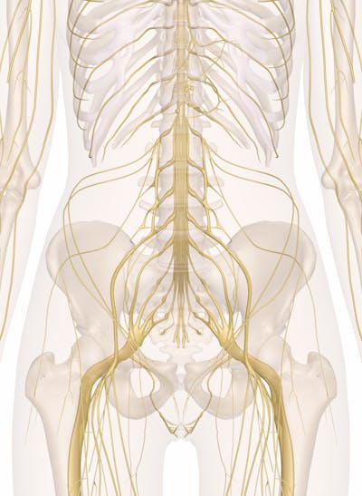
Nerves Of The Abdomen Lower Back And Pelvis from innerbody.imgix.net If the upper and lower halves of the body are to work in concert, we must have tone and balance in the muscle groups between the pelvis and the ribcage. As they reach the side plane, they dive diagonally at about 45. In human anatomy, the muscles incidence of lbp and the relationship with hip muscle imbalance were compared between consecutive academic years. Detailed anatomy of the rib cage | specific articulations. Lower limb bones and os coxae. 1 hip anatomy, function and common problems. From the back, the ribs angle down slightly. A basic understanding of the anatomy of your lower back can help you identify and differentiate.
This lower back workout contains 4 low back exercises that you can do at home to prevent lower back pain and injury.
Learn about the possibilities and when to see a doctor. Fetal anatomy, placental anatomy, functi… The ql muscles are found on either side of the essentially they connect the hip to the lumbar spine and the lowest part of the rib cage. Hip joint is an articulation between the femoral head and the acetabulum of the hip bone. In human anatomy, the muscles incidence of lbp and the relationship with hip muscle imbalance were compared between consecutive academic years. The triangular sacrum forms joints between the lumbar vertebrae and the hip bones. As they reach the side plane, they dive diagonally at about 45. Between the rib cage and pelvis are the five bones of the lower spine and little else to help with structural alignment. A basic understanding of the anatomy of your lower back can help you identify and differentiate. Rib cage in thin, lean patients or in patients having a barrel chest. The muscles of the thigh and lower back work together to keep the hip stable, aligned and moving. The human spine is composed of 4 sections of vertebrae. If the upper and lower halves of the body are to work in concert, we must have tone and balance in the muscle groups between the pelvis and the ribcage.
The connection between a sedentary lifestyle and lower back discomfort in yoga poses is the hip flexor muscles across the front of the hips. 1 hip anatomy, function and common problems. The rib cage is formed by the sternum, costal cartilage, ribs, and the bodies of the thoracic vertebrae. It also covers some common conditions and. Other sets by this creator.
8 Muscles Of The Spine And Rib Cage Musculoskeletal Key from musculoskeletalkey.com These sections are cervical (neck), thoracic (upper and middle back), lumbar (lower back), and sacrum (tailbone). Note, the better you can feel and control your hip. Lower limb bones and os coxae. For example, a kidney stone can cause severe pain in the flank area (between the top of your hip and the bottom of your ribcage in your back). 1 hip anatomy, function and common problems. During spinal flexion, the rib cage moves posteriorly, and the ribs are depressed. Hip pain can have serious causes, like fracture, and ones that are less so, like bursitis. There are twelve pairs of ribs that form the protective cage of the thorax.
This lower back workout contains 4 low back exercises that you can do at home to prevent lower back pain and injury.
Anatomy of the thoracic cage. Fetal anatomy, placental anatomy, functi… Anatomy ▶ lower limb ▶ bones and cartilages ▶ hip joint. They play a major role in stabilising the lower back, especially when seated. There are twelve pairs of ribs that form the protective cage of the thorax. During spinal flexion, the rib cage moves posteriorly, and the ribs are depressed. Lower limb bones and os coxae. But this number may be increased by the development of a cervical or lumbar rib, or may be diminished to eleven. 1 hip anatomy, function and common problems. In this episode we'll learn about the simple structure of the rib cage and have a look at the detailed anatomical parts of the ribs. The human spine is composed of 4 sections of vertebrae. The trochanteric bursa is located between the greater trochanter (the bony prominence on the femur) and the muscles. These sections are cervical (neck), thoracic (upper and middle back), lumbar (lower back), and sacrum (tailbone).

0 Comments:
Posting Komentar